Fowl pox is a slow-spreading viral disease in chickens characterized by lesions on unfeathered skin and/or the mucous membranes of the mouth, larynx, and trachea. It is caused by avian poxvirus, which exists in several strains, including
fowl poxvirus (affecting chickens and turkeys), pigeon poxvirus (affecting pigeons), and canary poxvirus (affecting various wild birds). Each strain is species-specific, meaning chickens cannot contract pigeon poxvirus, and vice versa.
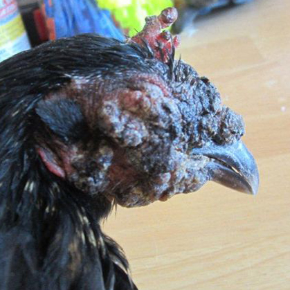
Forms of Fowl Pox
Fowl pox occurs in two forms: the dry (cutaneous) form and the wet (diphtheritic) form.
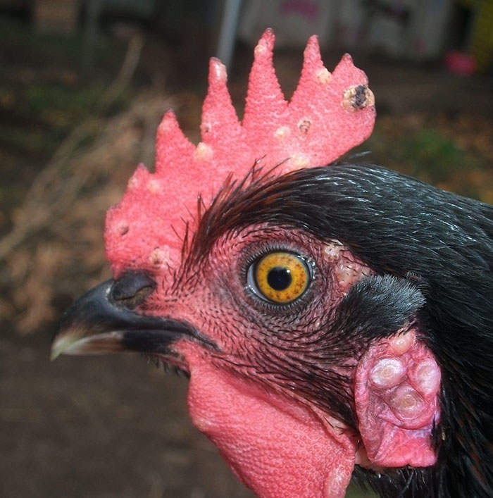
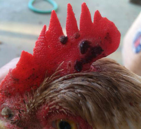
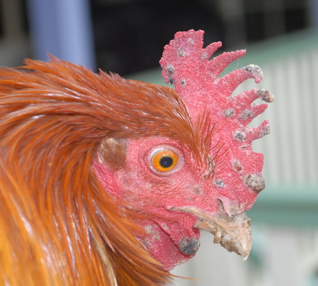
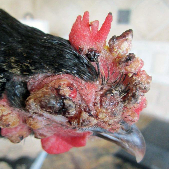
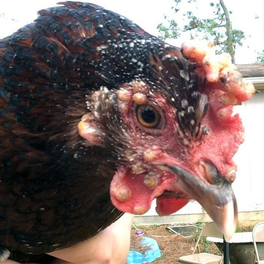
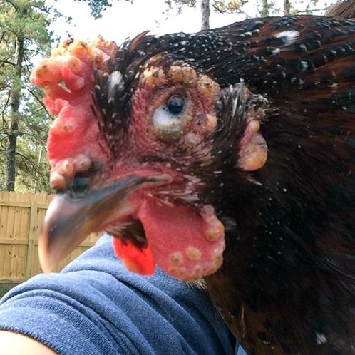
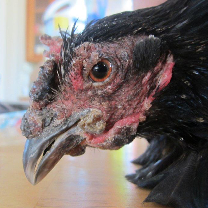
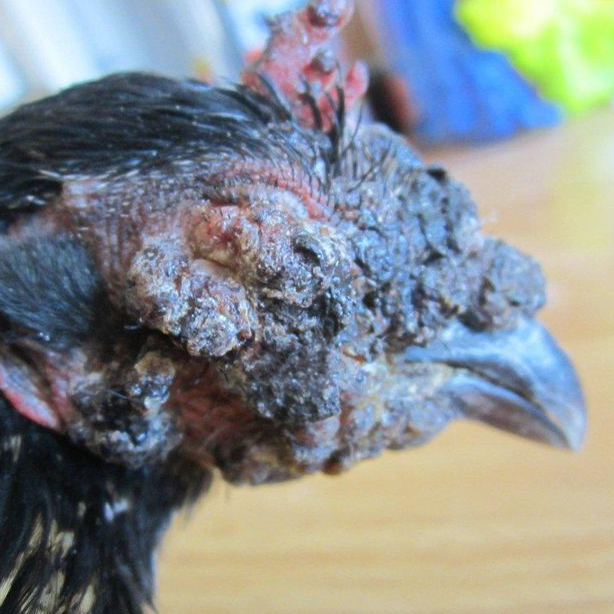
Dry (cutaneous) form
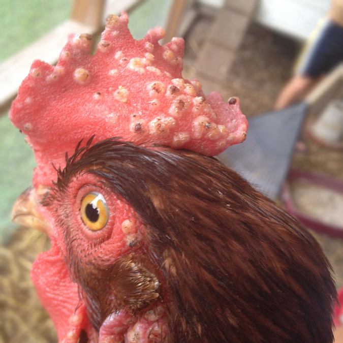
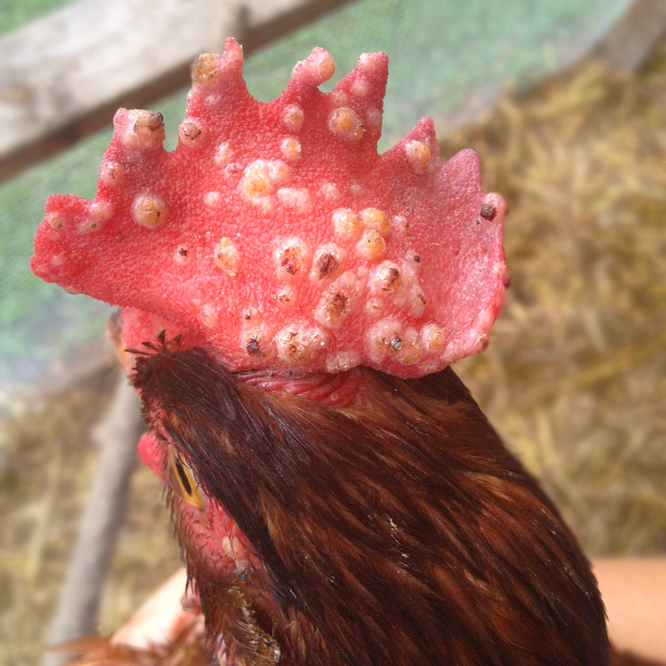
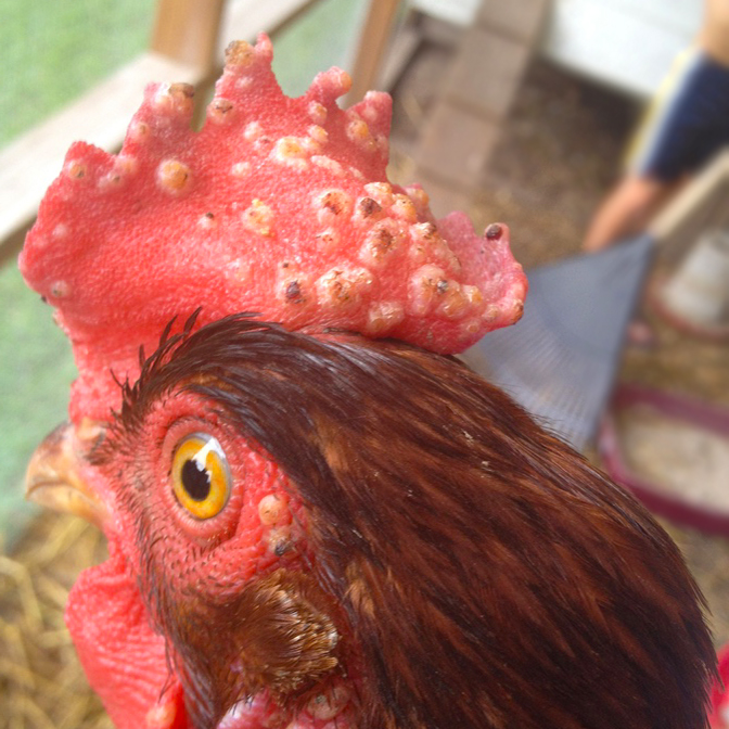
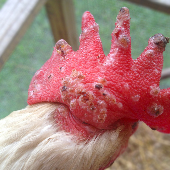
This is the most common presentation. It causes small, wart-like lesions on unfeathered areas such as the comb, wattles, face, eyelids, legs, and feet. These lesions begin as small yellow bumps that gradually enlarge and darken into rough, brown scabs. Scabs typically persist for 2–4 weeks before falling off, leaving smooth scar tissue.
These scabs contain active virus and are highly infectious. When lesions develop near the eyes, chickens may initially show mild irritation that can progress to swelling, ulceration, and closure of the eyelids due to discharge or crusting. During this stage, birds are more susceptible to secondary infections, so keeping lesions clean is important.
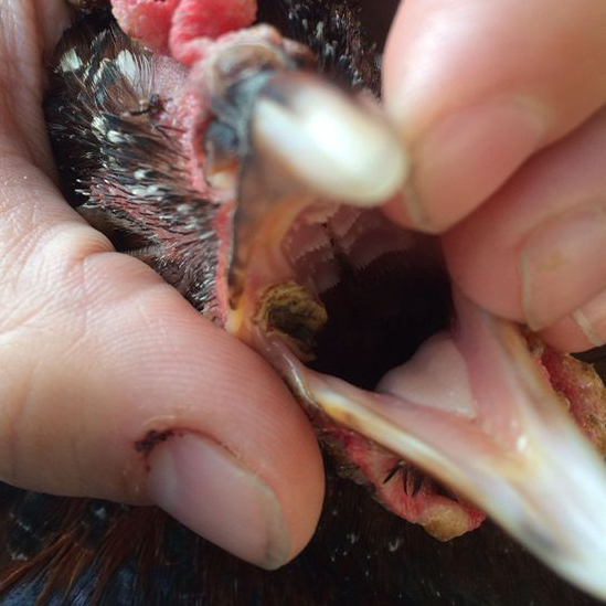
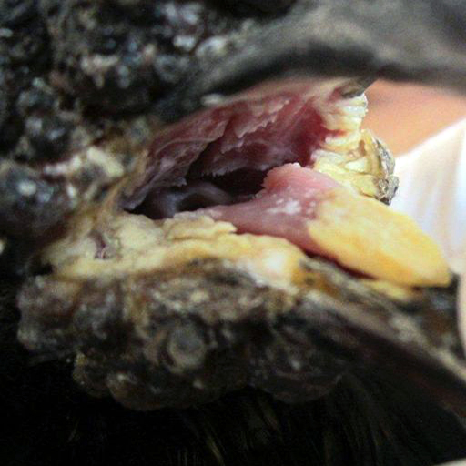
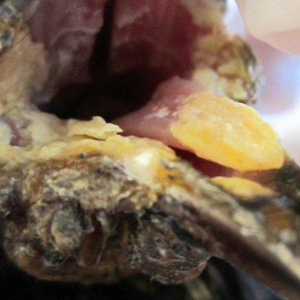
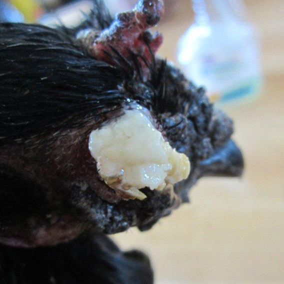
Wet (diphtheritic) form
This more severe form is associated with higher mortality. It causes yellow, cheese-like plaques (canker lesions) to form in the mouth and throat. These lesions may begin as small white nodules that merge into larger patches. When present in the upper digestive or respiratory tract, they can interfere with eating, swallowing, and breathing, leading to reduced feed intake and respiratory distress.
Transmission
Fowl pox is commonly spread by biting insects, especially mosquitoes, as well as by introducing infected birds into the flock. After feeding on an infected bird, mosquitoes can carry and transmit the virus for up to 8 weeks, potentially infecting multiple chickens.
Within a flock, the virus spreads through:
- Broken skin or mucous membranes: Often due to pecking or fighting.
- Dried scabs: which can remain infectious in the environment for months or even years.
Diagnosis
Diagnosis is based on flock history, clinical signs, and physical examination. Laboratory confirmation can be performed through virus isolation, fluorescent antibody testing, electron microscopy, or testing of scab or lesion samples.
Treatment
Fowl pox is typically self-limiting, meaning it resolves on its own over time. Treatment focuses on supportive care, including maintaining good hygiene and monitoring for complications. Antibiotics may be required if secondary bacterial infections develop.


], via Wikimedia Commons")
], via Wikimedia Commons")


]")
]")
]")


