Veterinary advice should be sought from your local veterinarian before applying any treatment or vaccine. Not sure who to use? Look up veterinarians who specialize in poultry using our directory listing. Find me a Vet
Squamous Cell Carcinoma

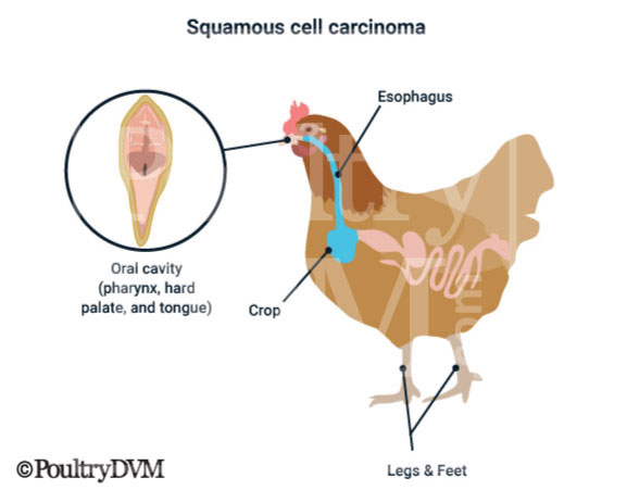
Squamous cell carcinoma (SCC) is a malignant tumor (cancer) that originates from squamous cells of the skin epithelium or digestive mucosa. Squamous cell carcinoma is one of the most common types of skin tumors that affect chickens. They can occur anywhere on the chicken's body but most common inside the oral cavity (pharynx, hard palate, and tongue), esophagus and uropygial gland. They can also occur on the beak, choana, wings, legs and feet.
Squamous cell carcinoma found on chickens
Clinical signs presented by chickens with squamous cell carcinoma vary depending on the location of the tumor. When it develops in the mouth or esophagus the most common signs include reluctance to eat, or reduced appetite, weight loss, and difficulty swallowing. Some birds may make wry neck like movements with their head and neck following consumption of food. There may be a visible mass in the oral cavity, or a palatable one along the outside of the bird's neck, in the area of the esophagus.

Tumors in the crop will often result in crop dilation, regurgitation, and vomiting. When they develop on the feet, legs, uropygial gland or wings they appear as ulcerated lesions or masses in the skin. SCC should be included in the differential diagnosis of integumental chronic ulcerated wounds, such as bumblefoot. Since bumblefoot is usually bilateral, if just one foot is affected the possibility of an ulcerated SCC should be considered.

Clinical Signs

Difficulty swallowing
Weight loss
Reduced appetite
Torticollis (wry neck) following feeding
Ulcerated soft tissue mass
Yellowish mass inside mouth
Weakness
Ataxia
Difficulty breathing
Regurgitation
Vomiting
Lethargy
Ulcer-like mass on one foot

Diagnosis

  • History
  • Clinical signs
  • Physical exam
  • Radiographs
  • Biopsy

Reported Cases

  • Case 1: Squamous cell carcinoma in a Chicken. A 4.5-yr-old rooster was presented for a “lump on the neck.” The bird exhibited dyspnea immediately prior to death. Necropsy revealed a crateriform lesion in the crop. Histologically, the lesion was diagnosed as an invasive squamous cell carcinoma, extending almost the entire thickness of the crop. Polymerase chain reaction and ultrastructural examination failed to identify a viral component. Early vascular invasion was identified. Ref
    Primary tumor site: cropSites of Metastases: none

  • Case 2: Squamous cell carcinoma in a Parrot. A case of metastatic squamous cell carcinoma in an African gray parrot caused by malignant transformation of an initial SCUD lesion that developed over a 2.5-year period. The right humerus was previously amputated to treat a squamous cell carcinoma, but it recurred in the skin 4 months prior to presentation. The bird presented on referral with pelvic limb paresis, and computed tomography showed a large, more right sided, paraspinal mass involving the right caudal coelom and effacing the lungs, ribs, and vertebral canal, resulting in the presenting neurologic signs. The patient was euthanized, and limited necropsy diagnosed the coelomic mass as a squamous cell carcinoma effacing the liver. Ref
    Primary tumor site: right humerusSites of Metastases: liver

  • Case 3: Squamous cell carcinoma in a Parrot. Squamous cell carcinoma of the rhinotheca was diagnosed in a 34-year-old timneh African grey parrot with a 2-year history of beak abnormalities. No evidence of metastasis or local invasion were found on results of radiographs or computed tomography scan. The bird was treated with surgical debulking and palliative megavoltage radiation therapy. After 4 radiation treatments, the affected tissue was necrotic and was debrided to reveal healthy granulation tissue. The bird died approximately 7 months after diagnosis and 4 months after cessation of radiation treatment. At the time of death, a small scab lesion remained at the left oral commissure, but no visible tumor regrowth was evident. A postmortem examination was not performed, however, and tumor recurrence could not be ruled out in this bird. Ref
    Primary tumor site: beakSites of Metastases: none

  • Case 4: Squamous cell carcinoma in a Cockatiel. A 20-year-old male cockatiel exhibiting dysphagia, regurgitation, and lethargy was diagnosed with invasive squamous cell carcinoma involving the crop. The neoplastic mass was closely associated with the right vagal nerve and was obstructing 80% of the crop lumen. Dysphagia appeared to be linked to nerve dysfunction resulting in ileus of the upper digestive tract. This is the first published report of squamous cell carcinoma in the crop of a psittacine species resulting in nerve dysfunction and possible vagal indigestion. Ref
    Primary tumor site: cropSites of Metastases: none

  • Case 5: Squamous cell carcinoma in a Budgie. A 2-year-old male budgerigar was referred to the Massey University Veterinary Teaching Hospital (MUVTH) after a 3-week history of regurgitation and vomiting. The budgerigar had been seen by the referring veterinarian on four occasions in the 3 weeks before referral. The bird was housed in an aviary with a 3-year-old male budgerigar and fed a commercial seed mix and millet sprays. There was no history of prior illness. Treatment before presentation to the MUVTH had consisted of 8 days of oral doxycycline (dose not reported). Ref
    Primary tumor site: cropSites of Metastases: none

  • Case 6: Squamous cell carcinoma in a Amazon. Squamous cell carcinoma of the distal portion of the esophagus was diagnosed, through use of contrast radiography and necropsy, in a 30-year-old Amazon parrot. The clinical signs were collapse, opisthotonos, and torticollis following feeding. Ref
    Primary tumor site: esophagus Sites of Metastases: none

  • Case 7: Squamous cell carcinoma in a Chicken. A mass of 7 cm in diameter, ulcerated, yellow, and interspersed with gray friable material was found in the esophagus. The bird had a history of lethargy, frequent loss of head and neck stability, difficulty eating, and difficulty swallowing. Large deep mass in the cranial cervical region, left eye was in miosis, with the pupillary margin of the iris slightly irrigular, and its ventral portion whitish. Ref
    Primary tumor site: esophagusSites of Metastases: none

  • Case 8: Squamous cell carcinoma in a Flamingo. A 32-year-old female American flamingo was presented with a squamous cell carcinoma of the middle digit of the right foot. No clinical, hematologic, or radiologic evidence of metastasis was present. Salvage amputation of the digit resulted in complete cure, whereas previous electrosurgery and radiation therapy were unsuccessful. Three years later, another squamous cell carcinoma was diagnosed in the middle digit of the left foot. The digit was also amputated. Seven months after the second amputation, the bird did not have any recurrence or signs of metastasis. Ref
    Primary tumor site: right footSites of Metastases: left foot

  • Case 9: Squamous cell carcinoma in a Chicken. Squamous-cell carcinoma was diagnosed on both legs of a 6-year-old pet Aracauna chicken. Grossly, the tumors were raised and horny with superficial ulceration. Histologically, the tumors were highly invasive, extending deep into the dermis, tendon sheaths, and periosteal tissues. There was no evidence of vascular invasion or metastasis to other organs. Ref
    Primary tumor site: legSites of Metastases: other leg

  • Case 10: Squamous cell carcinoma in a Chicken. Squamous cell carcinoma of the oropharynx and esophagus was diagnosed in an adult Japanese bantam rooster. Grossly, a cauliflowerlike mass with irregular edges was found involving the ventrolateral surfaces of the caudal portion of the oropharynx and cervical portion of the esophagus. The large volume of the mass almost occluded the lumen of the alimentary passage. Histologically, the tumor consisted of irregular cords of pleomorphic epithelial cells that showed a disorganized pattern of growth and invaded the adjacent tissues. Keratinized epithelial cells and moderate numbers of keratin pearls were readily observed. The mitotic index was low, and, although the tumor was locally invasive, there was no evidence of vascular invasion or metastasis. Ref
    Primary tumor site: oropharynx and esophagusSites of Metastases: none

  • Case 11: Squamous cell carcinoma in a Chicken. A backyard laying hen exhibiting muscular atrophy, dyspnea, and absence of egg production was analyzed for diagnostic insights. Gross findings revealed the presence of a large ulcerated mass with irregular edges involving the caudal part of the oropharynx and the cranial part of the esophagus, occluding the lumen of the esophagus and compressing the trachea. Small nodular lesions were detected also in the lungs. Histologically, both esophageal and pulmonary masses were characterized by nests of pleomorphic epithelial cells with squamous differentiation. The diagnosis was of squamous cell carcinoma of the esophagus with the uncommon feature of pulmonary metastasis. Ref
    Primary tumor site: oropharynx and esophagusSites of Metastases: none

  • Case 12: Squamous cell carcinoma in a Chicken. Squamous-cell carcinoma of the pharyngeal cavity was diagnosed in a 3-year-old Jersey black giant male chicken. Grossly, the carcinoma was round with irregular edges, yellow-tan, cauliflower-like with a crusty surface, and attached to the roof and sides of the pharyngeal cavity. Histologically, the surface of the mass was covered by a dense mat of necrotic mucosa and inflammatory cells. A broad front of neoplastic cords consisting of squamous epithelial cells extended into the lamina propria. There was no evidence of vascular invasion or metastasis. Ref
    Primary tumor site: oral cavity (pharynx)Sites of Metastases: none

  • Case 13: Squamous cell carcinoma in a Parrot. Squamous cell carcinoma of the infraorbital sinus was diagnosed in a 5-year-old Solomon Island eclectus parrot that presented with exophthalmos of the right globe and an ulcerative mass of 2-months duration at the right commissure of the beak. The mass was 2.5 × 2 × 2 cm and contained caseous exudate. The ulcerative lesion was surgically debulked, but the bird died and was submitted for necropsy. Histopathologic examination of the infraorbital sinus revealed squamous cell carcinoma with no evidence of metastasis. The trachea was heavily infiltrated with thick, septate fungal hyphae, and evidence of fungal angioinvasion was observed in the trachea and muscular layer of the crop. Ref
    Primary tumor site: sinusSites of Metastases: none

  • Case 14: Squamous cell carcinoma in a Chicken. A 2-year-old rooster local breed suffered from anorexia, emaciation with an abnormal large mass in the lateral side in the cervical region was attended to Veterinary Teaching Hospital belong to College of Veterinary Medicine, Mosul University. After physical examination a movable and friable mass was recognized in the cervical region, the case sends for surgical intervention. Gross examination of the removed mass showed a friable, pale, will capsulated with hemorrhagic areas. Microscopically, the mass included three types of tumors. Squamous cell thymic carcinoma characterized by the presence of keratin pearl. Lymphoma that characterized by atypical polymorphic lymphocytes, vesicular nucleus, and pyknotic nucleoli,while cytoplasm appears eosinophilic to granular with ill- define borders. Lipoma which appears as irregular shape of adipocytes that lack their nucleus, with scattered infiltration of pyknotic lymphocytes. This is the first pathological report of mixed tumor of the thymus gland in rooster that composed from squamous cell thymic carcinoma, lymphoma and lipoma. Ref
    Primary tumor site: skinSites of Metastases: none

  • Case 15: Squamous cell carcinoma in a Parrot. A 22-year-old male African grey parrot had episodes of chronic feather picking and self-mutilation for 10 years; it had a 5 cm diameter right axillary wound and a 2 cm left dorsal patagial wound. Initial treatment with azithromycin and wound management was unsuccessful. Biopsies of both masses indicated squamous cell carcinoma. The left patagial tumour was removed completely by electrocautery. Cisplatin was administered weekly into multiple sites on the right axillary tumour and it initially appeared to regress; however, the bird’s condition deteriorated after a month of treatment, and it was euthanased. The tumour was confirmed postmortem to be squamous cell carcinoma, which had invaded local tissues. The aetiology of the carcinoma may have been secondary to chronic focal trauma. Ref
    Primary tumor site: skinSites of Metastases: skin multiple sites (patagial and axilla)

Treatment

NameSummary
Supportive careIsolate the bird from the flock and place in a safe, comfortable, warm location (your own chicken "intensive care unit") with easy access to water and food. Limit stress. Call your veterinarian.
Surgical excision
Cryosurgery
Laser surgery
Radiation therapy
Electrochemotherapy
Photodynamic therapy
Topical medications
Supplemental Vitamin CHas anticancer effects on oral SCC tumors, to induce cell apoptosisJ Zhou et al., 2020
Manuka honeyFor birds suffering from oral SCC, application of Manuka honey has been shown beneficial for humans to reduce the effects of oral mucositis resulting from chemotherapy radiation.D Howlader et al., 2018

Support

Prevention

  • Ensure adequate vitamin A in diet.
  • Vitamin C has anticancer effects on oral SCC tumors to induce cell apoptosis.

Prognosis

Poor

Scientific References

Age Range

This is more commonly seen in older, adult chickens.

Risk Factors

  • Previous skin trauma
  • Previous respiratory illness or poxvirus lesions
  • Vitamin A deficiency
  • Chronic infection or inflammation